Recently, Prof. Wu Fugen of School of Biological Sciences and Medical Engineering of Southeast University and National Key Laboratory of Bioelectronics, cooperated with Prof. Chen Zhan of University of Michigan and synthesized ultrasmall and ultrabright organosilica nanodots with 100% photoluminescence quantum yield for the first time, and realized Long-Term Lysosome Imaging. Related results have been published in Nano Letters under the title of “One-Step Synthesis of Ultrasmall and Ultrabright Organosilica Nanodots with 100% Photoluminescence Quantum Yield: Long-Term Lysosome Imaging in Living, Fixed, and Permeabilized Cells”. The corresponding authors of the article are Prof. Wu Fugen and Prof. Chen Zhan. The first authors are SEU doctoral student Chen Xiaokai and Zhang Xiaodong.
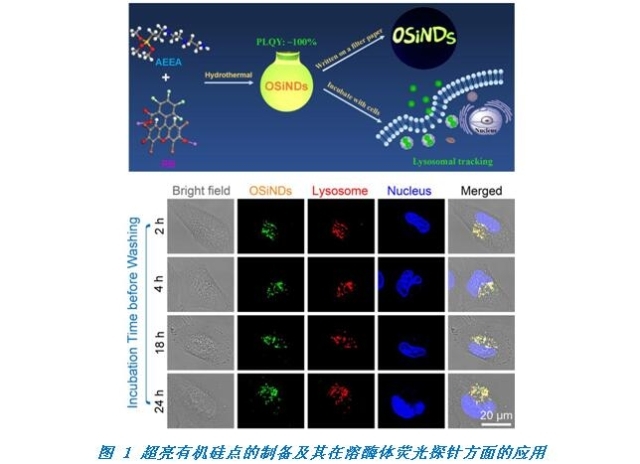
Water-dispersible nanomaterials with superbright photoluminescence (PL) emissions and narrow PL bandwidths are urgently desired for various imaging applications. Herein, for the first time, the team prepared ultrasmall organosilica nanodots (OSiNDs) with an average size of ~2.0 nm and ~100% green-emitting PL quantum efficiency via a one-step hydrothermal treatment of two commercial reagents (a silane molecule and rose bengal). In particular, the structural reorganization and halide loss of rose bengal during the hydrothermal treatment contribute to the ultrahigh quantum yield and low phototoxicity of OSiNDs. Owing to their low pH-induced precipitation/aggregation property, the as-prepared OSiNDs can be used as excellent lysosomal trackers with many advantages: (1) They have superior lysosomal targeting ability with a Pearson’s coefficient of 0.98; (2) The lysosomal monitoring time of OSiNDs is up to 48 h, which is much longer than those of commercial lysosomal trackers (<2 h); (3) They do not disturb the pH environment of lysosomes and can be used to visualize lysosomes in living, fixed, and permeabilized cells; (4) They exhibit intrinsic lysosomal tracking ability without the introduction of lysosome-targeting ligands (such as morpholine) and superior photostability; (5) The easy, cost-effective, and scalable synthetic method further ensures that these OSiNDs can be readily used as exceptional lysosomal trackers. The team expects that the ultrasmall OSiNDs with superior fluorescence properties and easily modifiable surfaces could be applied as fluorescent nanoprobes, light-emitting diode phosphor, and anticounterfeiting material, which should be able to promote the preparation and application of silicon-containing nanomaterials.
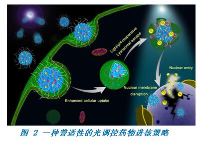
In another work, the team build a light-controlled nanoplatformfor direct nuclear delivery of molecular and nanoscale materials for the first time. Related results were published inJournal of the American Chemical Societywith the title “Development of a Light-Controlled Nanoplatform for Direct Nuclear Delivery of Molecular and Nanoscale Materials”, and the article was chosen as the cover paper of the journal. The corresponding authors of the article are Prof. Wu Fugen and Prof. Chen Zhan. The first authors are SEU post-graduate student Zhu Yaxuan and doctoral student Jia Haoran.
Research on nanomedicines has rapidly progressed in the past few years. However, due to the limited size of nuclear pores (9–12 nm), the nuclear membrane remains a difficult barrier to many nucleus-targeting agents. Here, the team report the development of a general platform to effectively deliver chemical compounds such as drug molecules or nanomaterials into cell nuclei. This platform consists of a polyamine-containing polyhedral oligomeric silsesquioxane (POSS) unit, a hydrophilic polyethylene glycol (PEG) chain, and the photosensitizer rose bengal (RB), which can self-assemble into nanoparticles (denoted as PPR NPs). Confocal fluorescence imaging showed that PPR NPs mainly located in lysosomes after cellular internalization. After mild light irradiation, however, PPR NPs effectively disrupted lysosomal structures by singlet oxygen (1O2) oxidation and substantially accumulated on nuclear membranes, which enabled further disruption of the membrane integrity and promoted their final nuclear entry. Next, the team selected two chemotherapeutic agents (10-hydroxycamptothecine and docetaxel) and a fluorescent dye (DiD) as payloads of PPR NPs and successfully demonstrated that this nanocarrier could efficiently deliver them into cell nuclei in a light-controlled manner. In addition to molecular compounds, the team have also demonstrated that PPR NPs could facilitate the nuclear entry of nanomaterials, including Prussian blue NPs as well as gold nanorods. Compared to traditional strategies for nuclear delivery, this highly controllable nanoplatform avoids complicated modification of nucleus-targeting ligands and is generally applicable to both molecular compounds and nanomaterials.
Southeast University is the first completing unite of the above two articles. The work was supported by grants from the National Natural Science Foundation of China, National Key Laboratory of Bioelectronics, the Natural Science Foundation of Jiangsu Province, Fundamental Research Funds for the Central Universities, Innovative and Entrepreneurial Talent Recruitment Program of Jiangsu Province, and the Six Talents Peak Project in Jiangsu Province.